When people fall ill, they go to a doctor’s surgery or a clinic, where medical professionals provide help. It may come as a surprise at first that physics also plays a not insignificant role in diagnosis and therapy. Since 2024, the bachelor’s degree programme in medical physics has been running at Bielefeld University in cooperation with Evangelisches Klinikum Bethel (EvKB), part of the University Medical Center OWL. With the subsequent master’s degree, students will be able to further qualify as medical physics experts starting in 2028.
The link between physics and medicine most likely began when the physicist Conrad Röntgen discovered the radiation, which was later named after him, more than 130 years ago. X-rays were soon used in examinations, but also in the treatment of patients with cancer and tuberculosis. And that was not the end of it: ‘Nowadays, medical physics methods are indispensable in both diagnostics and therapy,’ says Professor Dr Thomas Huser, who, with his working group ‘Biomolecular Photonics’ in the Faculty of Physics at Bielefeld University, researches the latest optical imaging. ‘They are used every day to prolong or save human lives.’
© Elena Berz
Medicine without physics? Unimaginable nowadays!
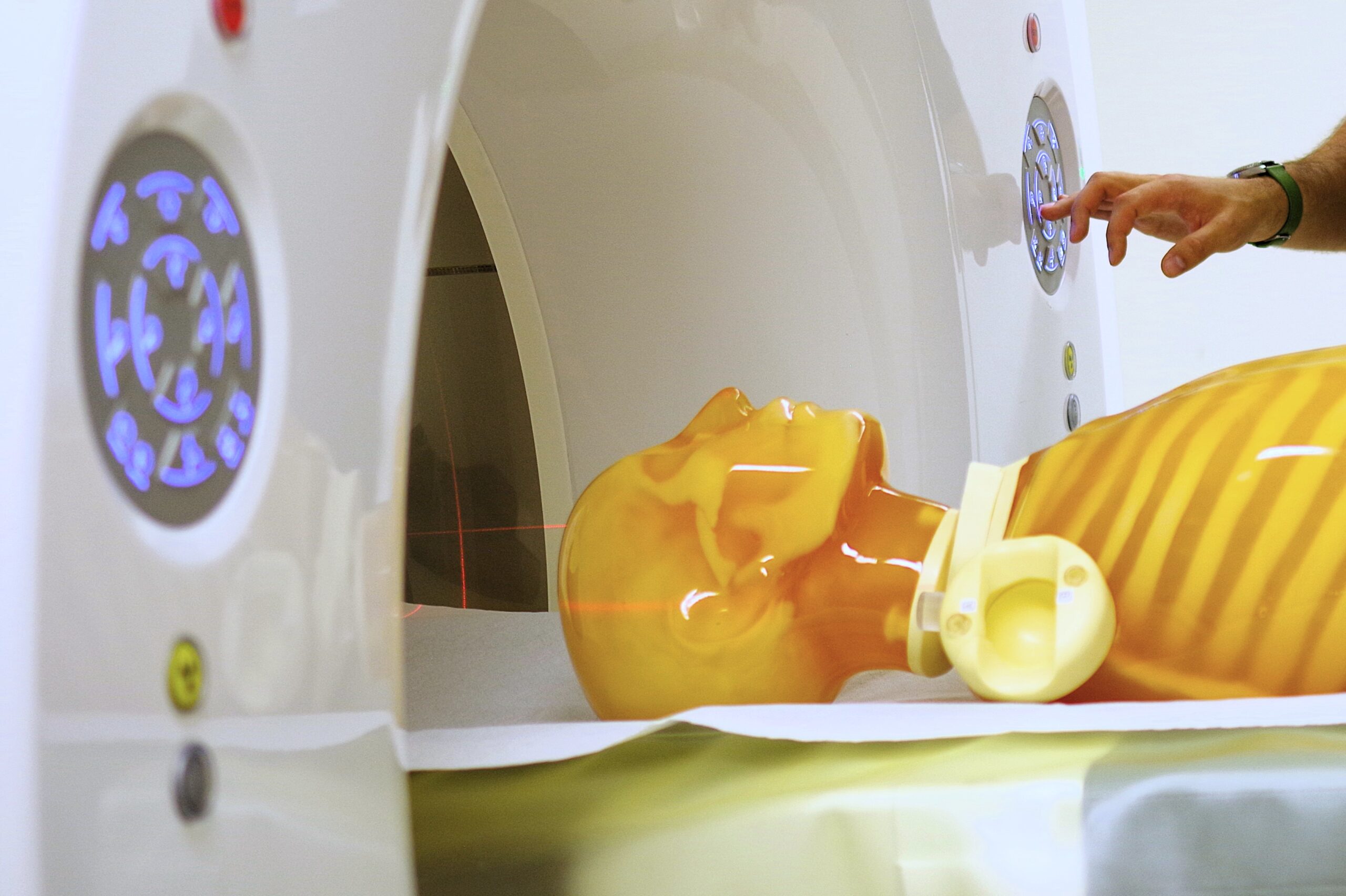
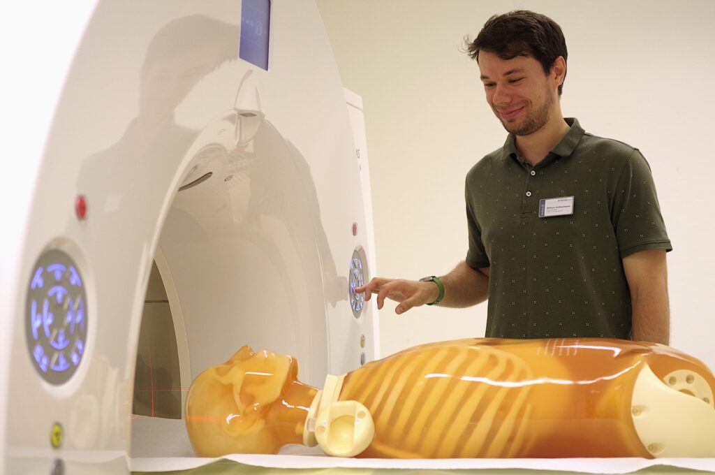
Thermometers, stethoscopes, blood pressure monitors, ventilators and electrocardiography (ECG) are just some examples of originally physical developments that were then integrated into medical routine. Magnetic resonance imaging (MRI) and computed tomography (CT) have also become indispensable in everyday clinical work. ‘Medical physics experts are needed to operate these and other devices, ensuring the safe and effective use of physical methods and technologies in the medical environment,’ explains Dr Volker Walhorn, a member of the ‘Biophysics and Applied Nanosciences’ working group led by Professor Dr Dario Anselmetti.
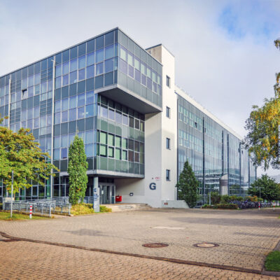
‘The aim of our new medical physics degree programmes,’ says Huser, ‘is to ensure that clinics are supplied with excellently trained medical physicists.’ For this purpose, Bielefeld University cooperates with Evangelisches Klinikum Bethel – ‘a win-win situation’, says Professor Dr med Günther Wittenberg, chief physician of the Institute for Diagnostic and Interventional Radiology and Paediatric Radiology, as well as head of the Institute for Medical Physics at EvKB.

© Elena Berz

© Elena Berz
Cooperation: teaching and research benefit
In order to be certified as a medical physics expert at the end of their studies, students must acquire the so-called professional competence, which means working at the clinic in a practice-oriented way. Thanks to the cooperation, this is easily possible in Bielefeld – unlike at other locations, where prospective medical physicists often have to find internship placements themselves: ‘We can enable students right here on site to put theoretical content from the university into practice – supervised by our radiologists and medical physics experts,’ says Wittenberg. That said, it is not only teaching but also research that benefit from the cooperation. ‘For us, this now opens up completely different possibilities in this area, also because we can use university resources – that is really top!’
Reducing radiation, optimising imaging
The central aim of medical physicists in hospital is to reduce patients’ radiation exposure – the keyword being radiation protection. ‘We are aiming to keep the radiation dose as low as possible while diagnosing with the greatest possible accuracy,’ says Günther Wittenberg. ‘For this purpose, research is being carried out to improve imaging so that we can move towards molecular imaging, which means that we being able to assess cells.’ In the future, this could possibly spare patients painful and risky procedures, such as tissue biopsies.
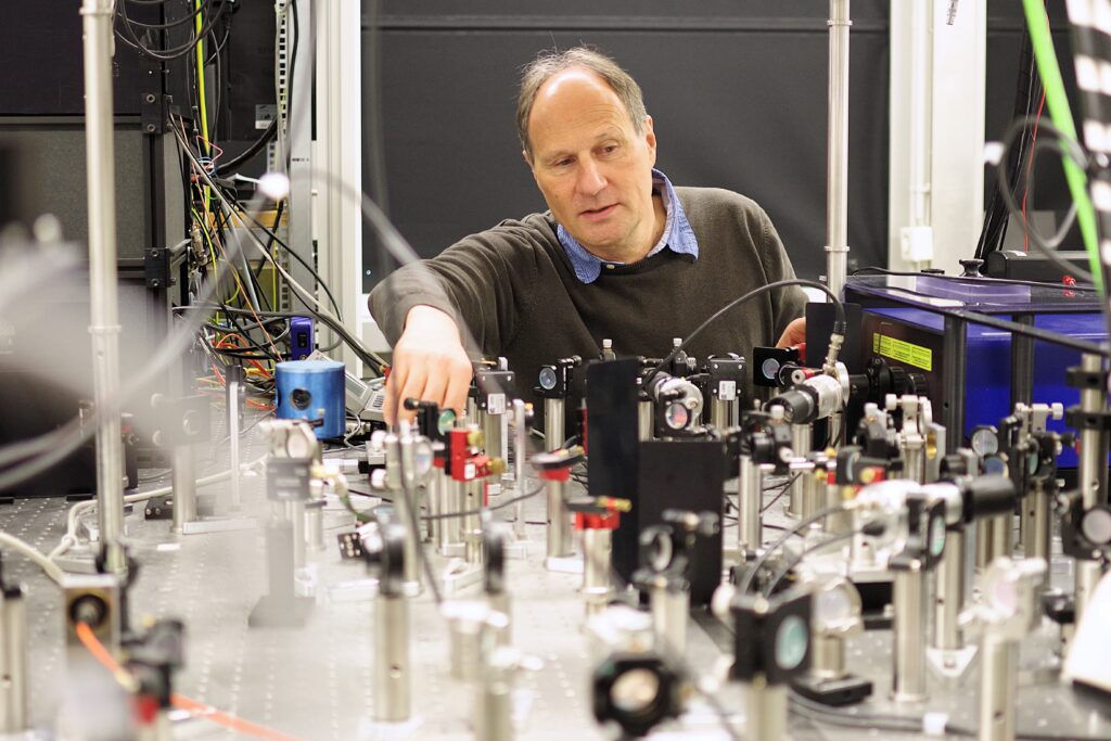
One of the people researching in this area is Thomas Huser. The physics professor has developed a new microscope that can make structures visible in cells that cannot be visualised using conventional methods. Prior to that, looking into such tiny organisms was only possible with electron microscopes. However, examining samples in this way is not only time-consuming and expensive – it also means that only dead cells can be observed. Huser’s microscope, by contrast, makes it possible to image living cells in real time.
By doing so, processes in microscopic structures can be identified and visualised, such as individual virus particles attacking cells and penetrating them. ‘Almost all viruses are significantlhy smaller than the resolution of advanced light microscopes, but thanks to our new, high-resolution methods we can capture this process and observe viruses on their journey into the cell,’ says Huser.
© Elena Berz
Real-time microscope offers new approaches to diagnosis
So far, Huser and his staff have mainly used the real-time microscope to investigate and help understanding basic cell biological processes. The filter system in liver cells, whose full investigation conventional microscopes lacked the necessary resolution, serves as one example for this. ‘We have recently made major advances here, and also with another organ, the kidney,’ says Huser. For the first time, the researchers have succeeded in imaging the structure of endothelial cells in the finest, highly porous blood vessels of the liver. These are crucial for the liver’s full function. The glomerulus, a filtering unit of the kidney, can now also be imaged at the highest resolution.
‘This kind of research advance potentially opens up new possibilities for diagnostics on biopsied tissue samples,’ says Huser. ‘Underfunction of the liver, for example, could be examined in real time instead of samples having to be checked as before in specialist laboratories using labour-intensive and costly electron microscopy.’ To make examinations with the real-time microscope possible in doctors’ surgeries and clinics, the startup project ‘Lightweaver’, which emerged from Huser’s working group, is working on automating and standardising processes such as the necessary tissue preparation – following that, the microscope could make the transition from the research laboratory into everyday medical use.
© Sarah Jonek
Watching life at work
Things are less application-oriented – but no less significant for this kind of research – in Professor Dr Dario Anselmetti’s working group. ‘We study the most fundamental molecular processes that make life what it is,’ says Dr Volker Walhorn. ‘Our aim is to understand how proteins function and how changes in their structure influence biological processes at molecular level.’
Using atomic force microscopy, the biophysicists investigate extremely small structures, some of them being a million times smaller than a millimetre. ‘This allows us not only to visualise surfaces, but also to represent the structure of individual molecules or cells. We therefore gain direct information about their composition,’ says Walhorn. Since structure and function are closely linked in biology and only two molecules that structurally fit together can interact with each other, the researchers also measure the binding forces between molecules and thus are able to map molecular processes. ‘Through this process, we gain a deeper understanding of how biological processes work at the smallest scale.’
Using these physical methods, Bielefeld researchers have been investigating inherited heart muscle diseases for more than 15 years, in cooperation with the Heart and Diabetes Centre in Bad Oeynhausen. The focus is on two proteins that ensure the mechanical connection between heart muscle cells. Put simply, on the molecular scale they function like Velcro. If these proteins are affected by genetic changes, this can cause serious heart muscle diseases. Although both the underlying defect in the genetic material and the clinical symptoms are usually known, the functional relationship is often unclear. ‘In other words: we know which molecule has changed, but often do not fully understand how this change disrupts the function of the heart muscle cells,’ explains Walhorn.
This knowledge gap is where the research of the Bielefeld scientists comes in. ‘We are watching life at work: by analysing the molecular processes that ensure heart muscle cells remain stably connected and the heart can beat reliably, we can understand how pathological changes arise. This understanding is crucial for the further development of diagnostics and therapy.’ After all, only those who understand how a system works in detail can reliably identify and rectify errors.
Studying medical physics
Ever since the winter semester of 2024/25, Bielefeld University has been offering the bachelor’s degree programme in medical physics as part of a three-pillar model in cooperation with Evangelisches Klinikum Bethel (EvKB), University Hospital OWL of Bielefeld University at the Bielefeld-Bethel campus. This model ensures a comprehensive and fully-fledged university education in physics, supplemented by appropriate minor and elective subjects with clinical medical physics content and basic knowledge of medicine.
Starting from the year after the next, Bielefeld University will also offer the opportunity to pursue a master’s degree in medical physics. Graduates have a wide range of career options open to them: as medical physics experts, they ensure the proper and safe functioning of systems in clinics with radiological facilities. Alternatively, their expertise is also in demand at manufacturers of medical devices and radiation protection equipment, or at authorities such as the Federal Office for Radiation Protection. With this degree, graduates can also go into research.
The application deadline for the coming winter semester ends on 15 July 2026.